High-Resolution Imaging of Fungi and Microorganisms Inside a Biosafety Cabinet
- Industry
- Budget
- Customer
Boost Productivity, minimize Costs! Discover how GOKO’s microscopes have unlocked new possibilities.
Strengthening Biosafety Measures! Clear and Magnified Observation of Fungi and Microorganisms Inside a Biosafety Cabinet Without Opening the Petri Dish
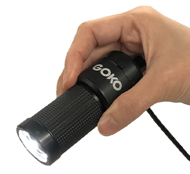
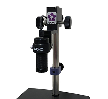
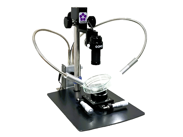
By introducing the Multi-Distance Scope GOKO EV-6HD, researchers can now observe fungi and other microorganisms with high clarity inside a biosafety cabinetーwithout the need to open the petri dish.
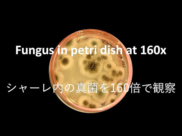
Traditionally, identifying mold required specimen preparation and approximately two weeks of incubation. However, with the GOKO EV-6HD, mold cultured overnight in a petri dish can be magnified and evaluated at an early stage.
This technology ensures safety while enabling rapid identification, significantly reducing the workload of laboratory technicians. By reinforcing biosafety measures, it also enhances research productivity in microbiology.
High-Resolution Imaging Comparable to ¥8.5 Million-Class Equipment
The Multi-Distance Scope GOKO EV-6HD delivers top-tier performance at an budget-friendly price. Researchers have even commented that its imaging quality is "comparable to an ¥8.5 million-class magnification device."
With over 70 years of optical expertise, GOKO provides unparalleled clarity and precision in magnified imaging.
High-Performance Measurement Software Empowering Scientific Research: GOKO Measure Plus
Quantifying and visualizing observation results is essential for generating reliable scientific data.
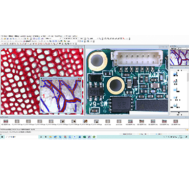
GOKO Measure Plus, GOKO's proprietary image measurement software, enables advanced analysis and precise measurement on high-resolution microscopic images ー all through an intuitive interface. It dramatically enhances research productivity and reproducibility.


Key Features:
- Smooth, intuitive measurement of dimensions, lengths, areas, and more using simple mouse operations.
- Automatic area selection, allowing rapid measurement of area, area ratio, and perimeter from auto-detected regions.
- Overlay and background subtraction functions let users compare live images with still images or detect differences between two static images with ease.
- Click-based object counting simplifies quantification of microorganisms, cells, and other small entities.
- Excel integration enables one-click transfer of both images and numerical data for seamless documentation and sharing.
- Scale display instantly visualizes object sizes in real time, enhancing clarity during observation.
These powerful features allow researchers to evaluate visual data both qualitatively and quantitatively, improving the reliability of research outputs and streamlining reporting, publication, and internal communication.
Compact and Portable for Flexible Use
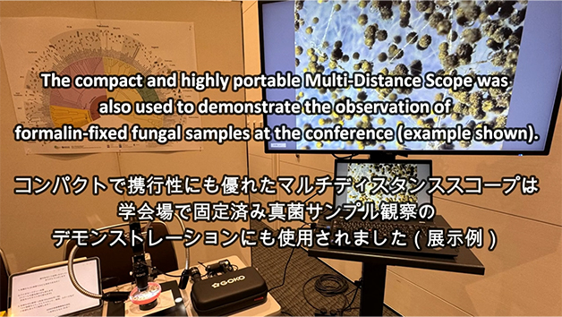
The GOKO EV-6HD has been praised for its compact and highly portable design, allowing for flexible use in various observation environments.
For example, it has been successfully used at academic conferences for live demonstration exhibits, where a large audience could observe magnified specimens in real time.
Fungus in petri dish at 160x
Additionally, the device can be connected to iPads (Type-C), Android smartphones, and tablets for seamless image observation and
sharing.
With remote collaboration apps like Zoom, real-time remote verification is also possible.
使用機種